
Use case · Biotransformation
Metabolite identification
Rank metabolite and conjugate hypotheses from isolated or enriched NMR datasets with explicit residual uncertainty.
- Starting point
- Isolated or enriched metabolite NMR from incubations or biofluids
- Typical input
- 1H and 13C, solvent (typically D2O or DMSO-d6), plus parent structure or formula
- Spectra output
- Ranked metabolite and biotransformation candidates with per-shift residuals
- Chemist's call
- Overlay authentic standard, confirm regioselectivity by 2D, or keep provisional
Who this job is for and why it stalls
Drug-metabolism (MetID) and metabolomics scientists use NMR when LC-MS alone cannot lock down regioselectivity — such as distinguishing which position on an aromatic ring underwent phase I hydroxylation, or confirming the attachment site of a glucuronide or sulfate conjugate.
The bottleneck is that biochemical candidate spaces are full of isobaric lookalikes and endogenous background compounds. Relying on a 1H database match without checking 13C shift residuals across the modified site can burn days chasing an endogenous lookalike or the wrong regioisomer.
How the Spectra workflow runs
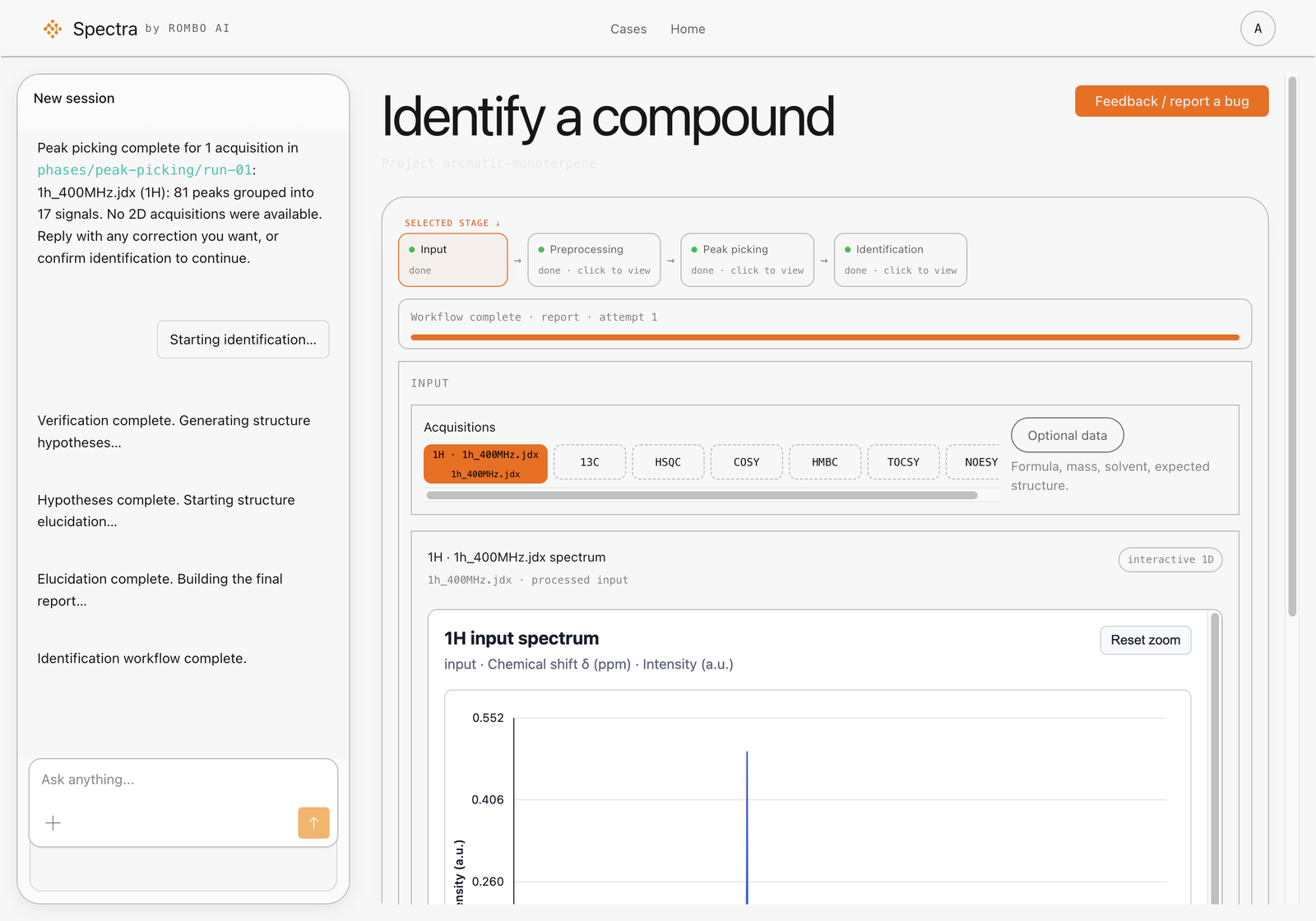
- Upload enriched metabolite 1H and 13C — Submit the NMR of the isolated or concentrated metabolite in
D2O,DMSO-d6, orCD3OD, and include the parent xenobiotic structure or molecular formula when available. - Rank regioisomers and biotransformation candidates — Spectra retrieves candidate structures and compares predicted versus observed chemical shifts across both the parent core and the modified positions.
- Evaluate carbon residuals at the site of metabolism — Use 13C RMSD and atom-level residuals to discard isobaric analogues whose carbon shifts conflict with the observed oxidation, dealkylation, or conjugation site.
- Inspect unexplained signals — Check unassigned peaks to see whether a conjugate moiety (such as a sugar or amino-acid fragment) or co-isolated matrix component is present.
What to look for in the review
- Clear 13C separation between competing regioisomers: helps prioritize which synthetic metabolite standard to order or synthesize first.
- Core matches well, but extra aliphatic/anomeric peaks remain unexplained: points toward a phase II conjugate or co-eluting endogenous component that requires targeted 2D (HSQC/HMBC) confirmation.
Related: unknown compound identification, evidence and confidence, limitations.
Scope & limitations
Where this workflow stops
Spectra does not automate pathway mapping, quantitative metabolomics, or MSI identification levels. Raw biofluids with heavy protein/lipid overlap and 1H-only profiling mixtures are outside scope; the workflow is designed for isolated or enriched material with 13C data.
Chemist sign-off
Recommended validation
Validate top-ranked hypotheses with an authentic standard or reference-spectrum overlay plus orthogonal MS, following the reporting standards required by your study or regulatory team.