
Use case · Extracts & fractions
NMR dereplication
Use Spectra to test whether an extract or mixture component is already known before committing columns and instrument time to isolation.
- Starting point
- Concentrated fraction or extract with a recurring spin system
- Typical input
- 1H at minimum; 13C when concentrated; solvent (e.g. DMSO-d6, CD3OD); optional fraction context
- Spectra output
- Ranked known library members, close analogues, and unexplained residual peaks
- Chemist's call
- Stop isolation on known compounds or prioritize novel residuals
Who this job is for and why it stalls
Natural-product and mixture chemists frequently encounter a clean spin system in a fraction and need to answer one question before scaling up chromatography: is this molecule already known?
Dereplication is a library problem with chemical nuance attached. Exact-match spectral searches break down when the solvent shifts slightly, when a minor component overlaps, or when 13C is partially incomplete. When a library search returns an inconclusive partial match, teams often isolate the compound anyway — spending days of preparative LC and magnet time on known flavonols, coumarins, sugars, or fatty-acid derivatives.
How the Spectra workflow runs
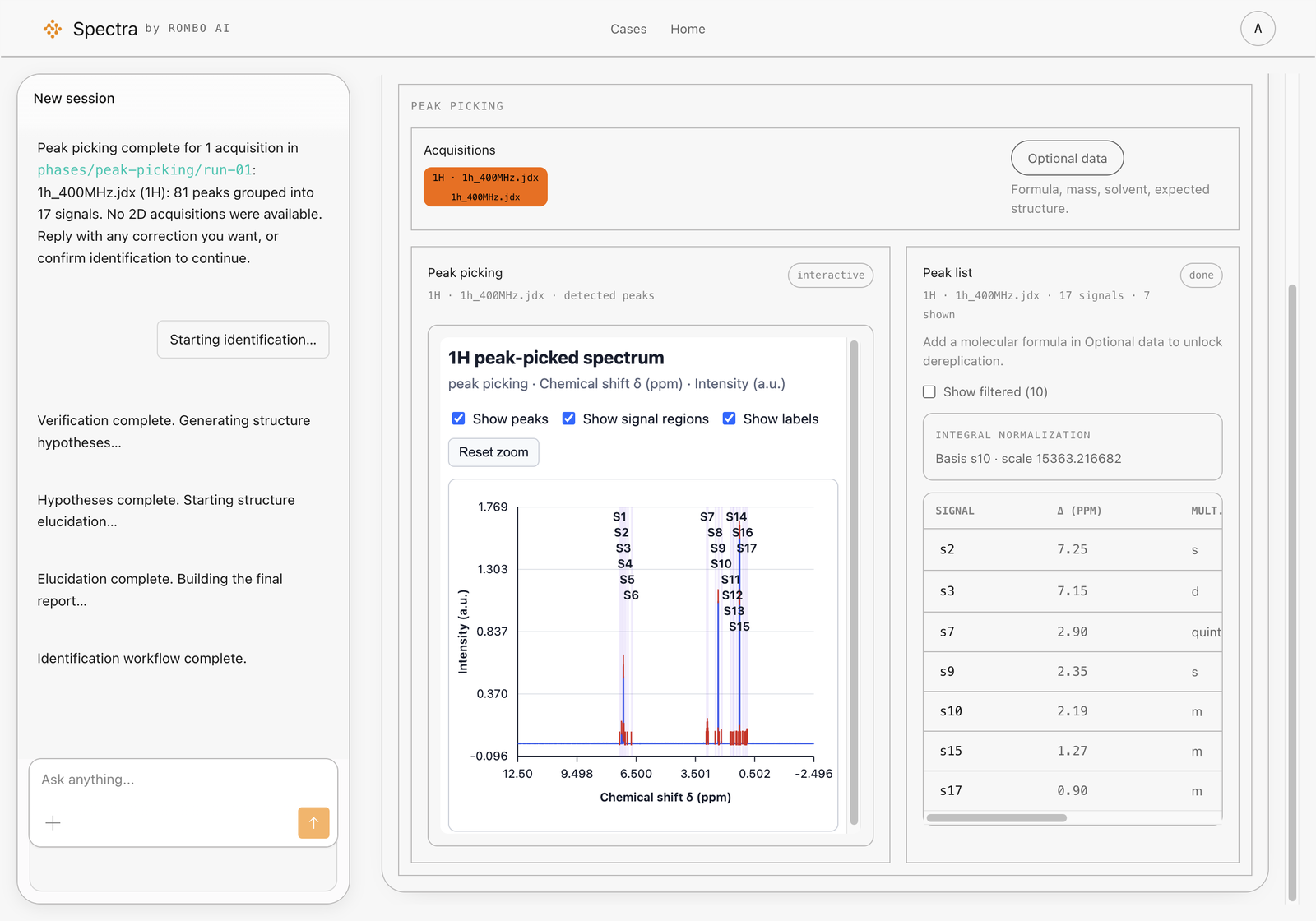
- Submit fraction NMR — Upload 1H and, whenever concentration permits, 13C data along with solvent and any molecular formula or mass context from LC-MS.
- Retrieve knowns and scaffold neighbours — Spectra searches across known structures and ranks candidates that fit the observed chemical shifts rather than relying on rigid peak-for-peak identity.
- Separate exact fits from scaffold analogues — Inspect the 13C RMSD and per-shift residuals. A low residual across the carbon skeleton points to a known compound you can set aside; a localized shift deviation flags a substitution difference worth isolating.
- Account for matrix leftovers — Review the unexplained signals list to distinguish co-eluting minor components from a genuinely different core structure.
What to look for in the review
- Low 13C residual across all diagnostic positions: strong evidence that the fraction’s main component is a known library member. Verify against an in-house standard and save the isolation column.
- High match on the core scaffold but a 3–6 ppm residual at one or two carbons: classic signature of a positional isomer, methylation pattern difference, or modified side chain. Keep in the isolation queue.
Related: natural-products research workflow, natural products and ingredient labs, sample analysis.
Scope & limitations
Where this workflow stops
A new analogue of a common scaffold can resemble the parent compound until 13C or 2D data are available. Dereplication is a triage filter, not a certificate of novelty or identity.
Chemist sign-off
Recommended validation
Overlay with an authentic standard or reference spectrum when available. When no standard exists, advance only the fractions whose 13C residuals remain unexplained by known library members.